Salivary Mucocele In Dogs And Cats
What is a salivary mucocele in dogs and cats?
A salivary mucocele in dogs and cats, also known as sialocele or salivary gland molecule, is swelling in the mouth caused by saliva leaking from the salivary glands or ducts and collecting in the soft tissue around them.
It’s most likely to occur in the mandibular gland, which is found below the jaw, and the sublingual gland, which is found underneath the tongue. You’ll see swelling under the skin and on the bottom of your pet’s mouth.
It can affect all dog breeds, but Poodles, German Shepherds, Australian Silky Terriers and Dachshunds are most susceptible. It can occur at any age and at any time and it can also occasionally affect cats, and is the most common salivary gland disease for both species.
Salivary mucocele symptoms in dogs and cats
The typical symptom of salivary mucoceles is an unstable swelling in your pet’s neck or mouth. Painless and usually soft, they tend not to impact your pet too much unless they grow large. If this happens, it can then result in symptoms such as:
- Difficulty eating
- Trouble swallowing
- Difficulty breathing
Where the salivary mucocele forms under the tongue (known as a ranula), it has the appearance of a bulge on the bottom of your pet’s mouth. If it grows large enough, you may be able to see it alongside the tongue, and your pet may experience:
- Difficulty closing its mouth
- Trouble eating
- Bleeding caused by damage to the mucocele when chewing
Pharyngeal salivary mucoceles are difficult to see, but you are likely to notice that your pet is:
- Having trouble breathing as the mass begins to obstruct its airway
- Experiencing shortness of breath
- Fainting
- Has difficulty swallowing
These symptoms must be assessed immediately by your vet so as to limit your pet’s distress and reduce the chance of death from severe respiratory distress.
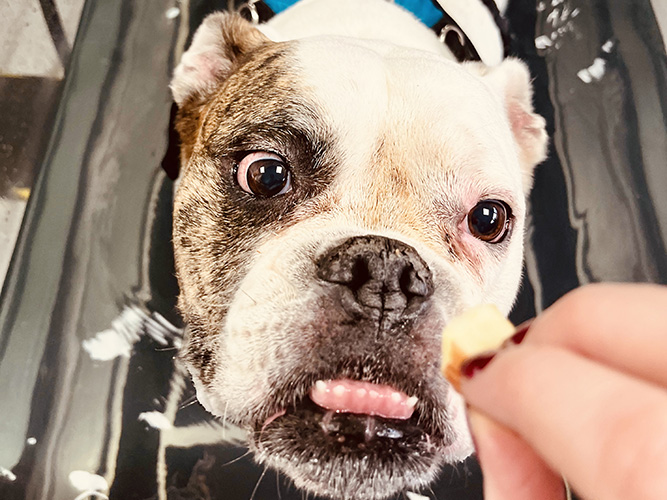
Though uncommon, you may also notice a swelling below your pet’s eye. This is most likely a zygomatic salivary mucocele, and it can cause your pet’s eye to bulge or appear larger than the unaffected eye. You might also see evidence of it on the roof of your pet’s mouth.
How is it diagnosed?
Your vet will carry out a thorough physical examination of your pet, which will involve looking in its mouth, and ask about its medical history.
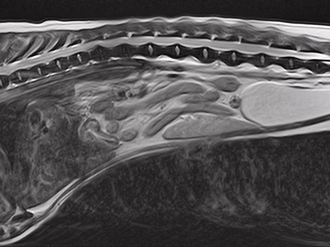
They may also need to carry out veterinary diagnostic imaging, such as a CT scan to determine where the swelling is located. Your pet will be sedated for this procedure.
This thorough examination is key, as there are other conditions that can cause swelling in the neck and mouth, including:
- Tumours
- Infections
- Abscesses
- Enlarged lymph nodes
- Cysts
In order to rule these conditions out, the most accurate diagnosis comes from aspirating the swelling or draining the fluid. Depending on the type of fluid that comes out, your vet will be able to determine the cause of the swelling. It’s most likely to be a salivary mucocele if the fluid is:
- Thick and stringy
- Clear
- Pale yellow
- Blood-tinged
This fluid can then be examined under a microscope, and assessed for bacteria or an elevated white blood count that would indicate an abscess or infection (known as sialadenitis).
If no bacteria is present, the fluid is determined to be saliva, and a positive diagnosis of a salivary mucocele in cats and dogs can be made.
If the swelling is hard or painful, your vet may suspect cancer. In which case, they will carry out an x-ray of the area to determine if that is the case. Your pet will be sedated for these procedures, so as to limit the amount of stress they may experience.
How is a feline and canine salivary mucocele treated?
There are many options that can be used to treat a salivary mucocele in dogs and cats, which are explained below:
Aspiration
Your vet will aspirate or drain the salivary mucocele, which will help to ease any pressure and irritation, and reduce the swelling. This is usually the first course of action, particularly if your pet is having difficulty eating, drinking, swallowing or breathing, and can provide quick relief.
If the area is infected, particularly inflamed or your pet is suffering with a lot of pain, your vet may prescribe antibiotics and anti-inflammatory medication to deal with the cause of the issue.
However, aspiration is only a temporary solution which may last a few weeks or months, but won’t solve the issue long term as the swelling can return. It also carries its own risk, as it can introduce bacteria into the area and make any subsequent treatment more difficult.
Surgical removal
The most effective treatment for a salivary mucocele is removal of the affected gland through soft tissue surgery. Your vet will not only remove the gland, they’ll also remove any diseased tissue.
Alternatively, where the salivary mucocele is caused by a damaged salivary duct, it may be possible for your vet to circumvent the issue and negate the need to remove the gland by creating a new opening in the salivary duct.
While surgical removal is a significant course of action, your pet has a number of salivary glands in its mouth, so removing one won’t impact it too much, and it won’t suffer from a dry mouth as a result.
Marsupialisation
Where the salivary mucocele occurs under the tongue, your vet may treat it with marsupialisation (cutting and stitching the mucocele so it can drain freely). Providing the fluid can drain away from the area, this procedure should enable the affected gland to heal.
What’s the prognosis?
The prognosis is excellent. Salivary mucoceles in dogs and cats should be cured following the surgery and your pet should go on to enjoy a normal life after treatment.
Providing the surgery is successful and every aspect of the affected gland has been removed (or sufficiently drained in the case of marsupialisation), the salivary mucocele shouldn’t return and your pet should recover well.
If your pet had surgery without complications, he/she can go home after recovery. Where marsupialisation has taken place, the drain will need to remain for several days.
If your pet has a resulting wound, the dressing will need to be replaced regularly too. If your pet’s wound is left open, you can help to keep the skin clean and encourage any fluid to drain by pressing a warm, damp towel to the area.
Your pet may develop a seroma, which is a pocket of fluid, at the site of their operation, but this can usually be left to heal on its own. It can also be drained, should your vet feel it’s necessary.
How long is recovery?
Once your pet is home, you will need to administer pain medication and any other medication, such as anti-inflammatories, that have been prescribed by your vet.
It’s important that your pet doesn’t scratch the area, and exercise will need to be restricted for three or four weeks post-surgery. However, once your pet has fully recovered and your vet is happy with their progress, normal exercise can be resumed.
You will need to attend follow-up appointments with your vet to make sure the area is healing, and to remove any drain that may have been used.
You should also closely monitor your pet during this time, and contact your vet immediately if there is any swelling, bleeding or difficulties with your pet’s breathing.
If you need any further advice on a salivary mucocele in dogs and cats, please don’t hesitate to contact us or fill in our veterinary referral form.